MEDICAL IMAGE DATA PROCESSING
w3
Project Overview
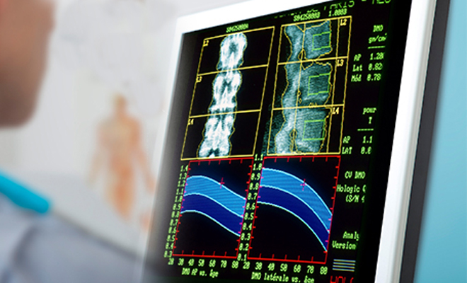
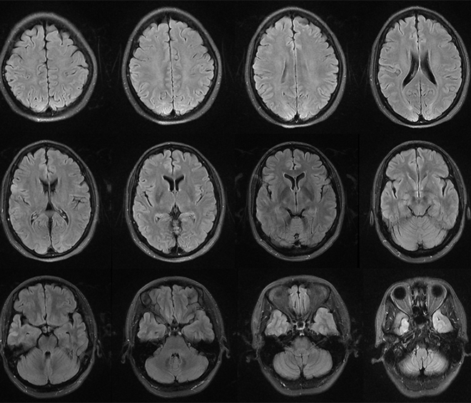
Medical image data processing is a key technology that involves in-depth analysis, interpretation, and extraction of effective information from medical images. These data may come from various medical imaging techniques, such as X-ray diffraction, computed tomography (CT), and magnetic resonance imaging (MRI). The main goal of medical image processing is to use advanced image processing and analysis algorithms to extract quantitative and qualitative information for disease diagnosis, treatment planning, and disease monitoring from raw images.
Medical imaging data processing plays a crucial role in the medical field. Firstly, it provides doctors with a powerful tool for accurately diagnosing diseases. By analyzing imaging data in detail, doctors can identify and locate abnormal structures, tumors or vascular lesions, and conduct precise quantitative evaluations, which is crucial for early detection and effective treatment of diseases. Secondly, medical image processing plays a crucial role in treatment planning and surgical navigation. By accurately processing and reconstructing imaging data, doctors can determine the most suitable treatment plan and accurately plan surgical steps, thereby reducing surgical risks and potential complications. In addition, medical imaging data processing is also an important means of monitoring disease progression and evaluating treatment effectiveness, providing valuable quantitative data for clinical research.
Scope of application
1586958963022fe877466966138f4bf31a8a400dd6b43
  • Image enhancement
    We use filtering, enhancement technology, contrast and brightness adjustment and other methods to improve image quality and visualization. This makes the details in the image clearer and easier for professionals to observe and interpret.
  • Segmentation and location
    Using the feature and density information of the image, we can accurately segment and locate the tissue, organ or abnormal structure in the image. This is very important for quantitative measurement, regional analysis and lesion detection.
  • 3D reconstruction
    By processing and fusing multiple slice images or plane photos and videos, we can generate three-dimensional models or volume rendered images to provide users with more comprehensive anatomical information.
  • Reconstruction and design of medical model
    Based on the segmented structure or organ, we use computer aided design (CAD) software for model reconstruction and design. This not only transforms medical image data into visual virtual models, but also transforms these virtual models into physical models through 3D printing technology.
  • Statistical shape model analysis
    We model and analyze a group of similar biological tissues, organs, cells and so on. Through the statistical analysis of a large number of samples, we can extract the variability and features of shape, and establish the probability model of shape variation, so as to reveal the statistical laws and characteristics of object shape.